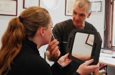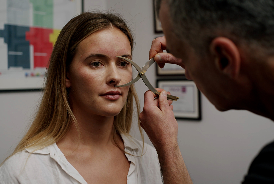
Anatomy-informed assessment for dermal fillers means mapping your unique facial structure, vascular anatomy, and tissue quality before treatment so that every placement decision is guided by your individual anatomy rather than a generic protocol.
- No two faces share identical anatomy, so a one-size-fits-all filler approach carries genuine safety risks.
- Vascular mapping using ultrasound technology identifies blood vessel locations specific to your face before injection.
- Skin quality, tissue thickness, and fat pad position all influence which filler product and technique is appropriate for you.
- Asymmetry assessment before treatment prevents the common mistake of adding volume to both sides equally when the underlying structure differs.
- A careful pre-treatment assessment is not a formality — it is the clinical step that determines whether your outcome is safe, natural, and proportionate.
An anatomy-informed assessment for dermal fillers maps your individual facial structure, vascular anatomy, skin quality, and tissue layers before any treatment begins. This process identifies anatomical risk zones, guides safe injection depth and placement, and ensures that hyaluronic acid-based dermal fillers enhance your natural features rather than alter them unpredictably.
Two people of the same age, with similar concerns, sitting in the same consultation chair. One has a high nasal bridge and thin skin over prominent cheekbones. The other has a broader, flatter midface with thicker subcutaneous tissue and a different vascular pattern running beneath the surface. Give them identical filler volumes in identical locations using identical technique, and you will get two entirely different outcomes, one of which could be genuinely dangerous.
This is the reality that makes anatomy-informed assessment not a premium add-on to filler treatment, but the clinical foundation without which safe, appropriate treatment cannot begin. If you have been researching dermal fillers and wondering why a careful pre-treatment assessment matters so much, this article explains exactly what that process involves and why your individual anatomy shapes every decision that follows. It connects directly to the broader argument explored in how dermal fillers restore facial structure rather than simply adding volume, where the structural basis of facial ageing is examined in detail.
An anatomy-informed assessment for dermal fillers maps your individual facial structure, vascular anatomy, skin quality, and tissue layers before any treatment begins. This process identifies anatomical risk zones, guides safe injection depth and placement, and ensures that hyaluronic acid-based dermal fillers enhance your natural features rather than alter them unpredictably.
What follows is a section-by-section look at why individual anatomy makes generic protocols genuinely risky, what a careful assessment actually involves, how asymmetry is evaluated before treatment, where the high-risk anatomical zones lie and why their location varies between individuals, and how all of these findings translate into a personalised treatment protocol.
—
Why Individual Facial Anatomy Makes a Generic Filler Protocol Genuinely Dangerous
The Anatomical Variables That Differ From Face to Face
Facial anatomy is not simply a matter of bone structure and skin colour. Beneath the surface, every face contains a layered arrangement of bone, periosteum, deep and superficial fat compartments, muscle, and a network of arteries and veins that varies considerably from person to person. The position of the facial artery, for example, does not follow a single fixed course. Research using cadaveric and imaging studies consistently shows that the path of key facial vessels, including the angular artery and the infraorbital vessels, varies in depth, lateral position, and branching pattern to a degree that makes assumptions about location genuinely unsafe.
Beyond vascular anatomy, the thickness and density of subcutaneous fat compartments differs between individuals and changes with age, weight fluctuation, and genetic factors. The medial cheek fat pad sits at a different depth in a patient with naturally thin skin compared to someone with denser subcutaneous tissue. The retaining ligaments that anchor facial soft tissue to deeper structures vary in their strength and position, influencing how tissue moves with age and how filler behaves once placed.
Bone structure introduces further variation. Orbital rim projection, malar eminence height, mandibular angle definition, and chin projection all influence where volume loss is most visible and where structural support is most needed. Two patients presenting with similar surface-level concerns, perhaps flattening of the midface and early jowl formation, may have arrived at that appearance through entirely different anatomical pathways. One may have significant malar bone resorption; the other may have primarily fat pad descent with relatively preserved bone. The filler approach for each is different.
How Anatomical Variation Changes Injection Depth, Volume, and Product Choice
Injection depth is not a stylistic choice. It is a clinical decision driven by the anatomy of the specific zone being treated in the specific individual being treated. Placing filler too superficially in a region where the skin is thin can cause visible irregularity, a bluish discolouration known as the Tyndall effect, or prolonged swelling. Placing it too deeply in a zone where a vessel runs at an unexpected depth creates vascular risk.
Product selection follows the same logic. Hyaluronic acid-based dermal fillers are formulated with varying degrees of cross-linking, which determines their firmness, lift capacity, and tissue integration. A firmer, more cohesive filler placed too superficially in someone with thin skin may be visible or palpable. A softer filler used where structural lift is needed will not provide adequate support and may migrate. Skin thickness, tissue density, and the depth of the target plane all inform which product formulation is appropriate for which zone in which individual.
Volume decisions are equally anatomy-dependent. Overfilling a compartment that is anatomically small, or that sits adjacent to a facial nerve branch, can create distortion, asymmetry, or functional discomfort. The appropriate volume for a given area is not determined by a standard protocol. It is determined by the space available, the tissue characteristics, and the clinical goal, all of which require individual assessment before any syringe is prepared.
—
What an Anatomy-Informed Filler Assessment Actually Involves
Static and Dynamic Facial Analysis: Reading Movement as Well as Structure
A careful pre-treatment assessment begins with observation, and that observation extends well beyond the face at rest. Static analysis examines your facial proportions, the relationship between your upper, middle, and lower thirds, the position and projection of key bony landmarks, and the distribution of volume loss across your face. This gives a structural map of where support has diminished and where tissue has descended.
Dynamic analysis adds a layer that static examination cannot provide. Watching how your face moves during expression reveals the behaviour of the underlying musculature, the degree to which dynamic lines are contributing to your concerns, and whether certain areas of apparent volume loss are partly driven by repeated muscle contraction rather than structural change alone. This distinction matters because filler and botulinum toxin address different mechanisms, and the appropriate combination, if any, can only be determined by observing both states.
Dynamic analysis also reveals how skin quality and elasticity respond to movement. Skin that shows significant crepiness or laxity when animated may not respond to filler in the same way as firmer, more elastic tissue, and this observation feeds directly into the assessment of skin quality.
How Ultrasound Technology Supports Pre-Treatment Vascular and Tissue Assessment
Ultrasound technology has become an increasingly valued tool in aesthetic medicine assessment, not only for its role in identifying vessel location before injection, but for its broader diagnostic value in evaluating tissue layers, identifying previous filler deposits, and guiding clinical decision-making in complex cases.
Pre-treatment ultrasound assessment allows a practitioner to visualise the depth and approximate position of key vessels in the area being treated. Because vascular anatomy varies between individuals, this real-time imaging provides information that surface anatomy landmarks alone cannot reliably deliver. In high-risk zones such as the glabella, nasolabial fold, and periorbital region, knowing where a vessel runs in your specific anatomy is a meaningful safety consideration, not a theoretical one.
Ultrasound also reveals the depth of tissue planes and can identify the presence of previous hyaluronic acid filler that may not be clinically apparent on examination. This is particularly relevant if you have had treatment elsewhere, as undisclosed or forgotten filler can alter the tissue environment and influence both safety and outcome of further treatment.
Skin Quality Evaluation and What It Reveals About Filler Suitability
Skin quality assessment examines thickness, elasticity, hydration, and surface texture. These characteristics influence both the choice of filler formulation and the realistic expectations that can be set for any treatment.
Thin, photodamaged skin with reduced elasticity behaves differently under filler than thicker, well-hydrated tissue. In thinner skin, even correctly placed filler may show subtle surface irregularity, and the risk of the Tyndall effect is higher in superficial planes. Skin with significant laxity may not benefit from volume addition alone; in some cases, addressing skin quality through treatments such as microneedling with Dermapen 4 or skin booster bio-remodelling injections before or alongside filler treatment produces a more cohesive outcome.
VISIA skin analysis, which uses multi-spectral imaging to reveal subsurface changes including UV damage, pore structure, and pigmentation patterns, adds an objective dimension to the skin quality evaluation. Whilst VISIA is primarily associated with skin health assessment, the information it provides about the condition of your skin’s deeper layers is relevant to the overall picture of tissue quality that guides filler planning.
—
Assessing Facial Asymmetry Before Filler Treatment: What Most People Overlook
Natural Asymmetry Versus Acquired Asymmetry and Why the Distinction Matters
Every human face is asymmetrical. The degree varies, but no face is a perfect mirror image of itself, and most people, when shown an unflipped photograph of themselves, are mildly startled by the asymmetry they had not consciously registered. This natural asymmetry is not a flaw to be corrected. It is part of what makes a face recognisably yours.
The clinical distinction that matters is between natural asymmetry, which is structural and present throughout life, and acquired asymmetry, which develops over time as a result of differential ageing, habitual muscle use, dental changes, previous trauma, or prior aesthetic treatment. A natural asymmetry in jaw width, for example, may reflect underlying bone structure that has always been present. An acquired asymmetry in cheek volume may reflect the fact that one side of the face has lost fat pad volume more rapidly than the other, perhaps related to sleeping position, dental occlusion, or the pattern of bone resorption.
Distinguishing between these two types of asymmetry before treatment is essential because the clinical response to each is different. Attempting to correct a structural, bony asymmetry with soft tissue filler alone will not achieve the intended result and may create a different kind of imbalance. Understanding the origin of an asymmetry guides whether, where, and how much filler is appropriate on each side.
Why Treating Both Sides Identically Often Creates Rather Than Corrects Imbalance
This is one of the most important and most frequently misunderstood aspects of filler treatment. Because your two sides are not anatomically identical, applying the same volume of filler to the same anatomical point on each side will not produce a symmetrical outcome. The tissue environment on each side, including skin thickness, fat pad depth, and underlying bone projection, differs enough that identical treatment produces different results.
A careful assessment maps the specific differences between your two sides before any treatment begins. If your left malar eminence has slightly less projection than your right, the appropriate filler volume and placement on the left will differ from what is needed on the right. If one nasolabial fold is deeper than the other because of differential fat pad descent rather than a difference in skin quality, the treatment approach for each side needs to reflect that distinction.
The goal of asymmetry assessment is not to create perfect symmetry, which is neither achievable nor natural-looking, but to reduce the degree of imbalance in a way that respects the underlying structure of your face. Realistic expectations in this context are important, and what to expect from dermal filler outcomes and when explores this in practical detail.
—
High-Risk Anatomical Zones and How a Careful Assessment Reduces Your Risk
The Vascular Danger Zones of the Face and Why Their Location Varies Between Individuals
Certain areas of the face carry a higher risk of vascular complications during filler treatment. These include the glabella, the nasolabial fold, the nose, the periorbital region, the temples, and the lips. The risk in these areas arises from the proximity of arterial vessels whose inadvertent injection can cause tissue ischaemia, and in the most serious cases, vision changes or skin necrosis.
What makes these zones genuinely complex is that the depth and lateral position of the relevant vessels is not fixed. The facial artery, for example, takes a variable course across the midface. In some individuals it runs close to the nasolabial fold; in others it sits more laterally or more deeply. The supratrochlear and supraorbital vessels in the forehead region vary in their exit points from the bone. The angular artery near the nose varies in depth and distance from the alar base.
This variability means that knowing the general location of a danger zone is not sufficient for safe practice. Understanding where the relevant vessels run in your specific anatomy, informed by clinical assessment and where appropriate by pre-treatment ultrasound, is what allows a practitioner to adjust technique, injection depth, and product selection to reduce risk in your particular case.
How Pre-Treatment Mapping Changes the Clinical Approach to These Areas
Pre-treatment assessment of high-risk zones does not eliminate risk entirely, but it meaningfully changes the clinical approach in ways that matter for your safety. When a vessel is identified running at an unexpected depth or position, the practitioner can adjust the injection plane, choose a blunt cannula rather than a sharp needle, modify the volume used, or in some cases recommend against treating that specific area.
Ultrasound-guided assessment is particularly valuable in the periorbital and temple regions, where vessels run in close proximity to structures that are difficult to assess by surface examination alone. In the nose, where vascular complications carry a disproportionately high risk, pre-treatment imaging of the dorsal nasal artery and its branches informs whether treatment is appropriate and, if so, which technique minimises risk.
Beyond vascular mapping, pre-treatment assessment of high-risk zones includes evaluating the tissue characteristics of the area, its skin thickness, the depth of the target plane, and the presence of any previous filler that might alter the tissue environment. All of these factors feed into a clinical decision about whether, how, and with what product to treat a given zone.
—
How Your Assessment Findings Shape Your Personalised Treatment Protocol
Translating Anatomical Findings Into Filler Product Selection and Placement Strategy
Once your assessment is complete, the findings translate directly into a personalised treatment protocol. This is not a process of selecting from a menu of standard treatments. It is a process of matching the clinical characteristics of your anatomy to the properties of available filler formulations and the technical options for their placement.
A patient with significant malar bone resorption and good overlying skin quality may benefit from a firmer, more cohesive hyaluronic acid filler placed in the deep medial cheek fat compartment, providing structural lift that restores the support architecture of the midface. A patient with a similar surface presentation but thinner skin and a more superficial fat pad loss may require a softer formulation placed at a different depth to avoid surface irregularity.
The placement strategy, meaning the specific injection points, depths, and volumes for each area, is determined by the map built during assessment. Which zones will be treated in a single session, which may require staged treatment over multiple appointments, and which will be deferred until other concerns are addressed first: all of these decisions emerge from the assessment findings rather than from a predetermined sequence.
The assessment also determines the order of treatment. In some cases, addressing one area first changes the appearance of an adjacent area enough that the planned volume for the second zone needs to be revised. This kind of sequential, adaptive approach is only possible when the practitioner has a detailed understanding of your individual facial anatomy and how the different zones relate to one another.
When Assessment Findings Indicate That Filler Is Not the Right Approach
An anatomy-informed assessment does not always conclude with a filler prescription. There are circumstances in which the findings indicate that a different approach is more appropriate, and communicating this honestly is a core part of the diagnostic-first methodology.
Significant skin laxity without meaningful volume loss, for example, may not respond well to filler. Adding volume to tissue that lacks the structural integrity to support it can create heaviness or distortion rather than improvement. In such cases, addressing skin quality first, through bio-remodelling skin boosters, Dermapen 4 microneedling, or other modalities, may be a more appropriate starting point.
Where the degree of structural change is beyond what non-surgical treatment can meaningfully address, an honest assessment includes that conversation. Referral to a surgical colleague, where appropriate, is a clinical decision made in your interest, not a failure of the non-surgical approach. The assessment process exists to serve your wellbeing, not to find a justification for treatment.
Similarly, if previous filler is identified on ultrasound in an area where further treatment is planned, the protocol may include dissolution of that filler before new product is placed, ensuring that the tissue environment is appropriate and that the outcome of new treatment is not compromised by what was placed before.
—
Frequently Asked Questions
How long does an anatomy-informed filler assessment take?
A careful pre-treatment assessment typically takes between 30 and 60 minutes, depending on the complexity of your concerns and whether imaging tools such as VISIA or ultrasound are incorporated. The time invested in assessment directly influences the safety and appropriateness of any treatment that follows.
Can ultrasound really show where my blood vessels are before filler treatment?
Yes, diagnostic ultrasound can visualise superficial vessels in real time, including the depth and approximate course of key facial arteries in the area being assessed. It does not map every vessel with absolute precision, but it provides clinically meaningful information that surface anatomy landmarks alone cannot reliably deliver, particularly in high-risk zones.
Does my skin thickness actually affect which filler is used?
Directly, yes. Thinner skin requires softer filler formulations placed at appropriate depths to avoid surface visibility and the Tyndall effect. Thicker tissue with more subcutaneous depth can accommodate firmer, higher-lifting products. Skin quality assessment is a standard part of pre-treatment evaluation for this reason.
What happens if my face is quite asymmetrical? Will filler make it look worse?
Not if the asymmetry is assessed and accounted for before treatment. A careful pre-treatment evaluation distinguishes between natural and acquired asymmetry, maps the specific differences between your two sides, and uses that information to guide differential treatment. Applying identical volumes to both sides without this assessment is what risks worsening imbalance.
Is a VISIA skin analysis part of a filler assessment?
VISIA skin analysis is primarily a skin health diagnostic tool, and it is not a mandatory component of every filler assessment. However, the information it provides about subsurface skin condition and tissue quality is relevant to the overall evaluation, particularly when skin health concerns are part of the clinical picture alongside structural volume concerns.
—
The Assessment Is the Treatment, Begun
There is a tendency to think of the assessment as the step before treatment. In practice, the assessment is where treatment begins. Every observation made during a careful pre-treatment evaluation, whether about your bone structure, your vascular anatomy, your skin quality, or the specific pattern of asymmetry in your face, shapes what happens next in a way that no amount of technical skill can compensate for if that information is absent.
Your face is not a version of a standard facial template. It is a specific anatomical environment with its own structural characteristics, its own vascular geography, and its own history of change over time. The filler protocol that is appropriate for you reflects all of that specificity, and it can only be designed once that specificity is understood.
If you are considering hyaluronic acid-based dermal fillers and want to understand what a diagnostic-first assessment would reveal about your individual anatomy, we invite you to begin with a consultation at the practice. The conversation starts with your face, not with a treatment list.
Related Articles
- how dermal fillers restore facial structure
– Parent pillar establishes the structural rationale for filler treatment; linking here contextualises why individual anatomy assessment is the logical prerequisite to structural correction. - what realistic filler outcomes look like and when to expect them
– After explaining how assessment shapes the treatment protocol, linking to the realistic expectations article helps readers understand the timeline and nature of outcomes that follow an anatomy-informed approach.