Identifying whether your pigmentation is melasma, sun damage, or post-inflammatory hyperpigmentation is the single most important step before treatment, because each type responds differently and the wrong approach can make discolouration worse.
- Melasma, sun damage, and post-inflammatory hyperpigmentation look alike but behave very differently and require distinct treatment approaches.
- The distribution pattern of your pigmentation, where it appears on your face, is one of the most reliable early indicators of its type.
- Treating the wrong pigmentation type, such as using aggressive exfoliation on post-inflammatory marks, can deepen discolouration rather than improve it.
- Hormonal influences, UV exposure, and skin injury each trigger pigmentation through different biological pathways, which is why the cause matters as much as the appearance.
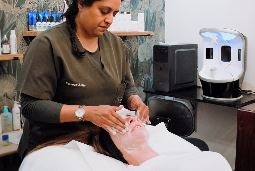
- A detailed skin assessment, including VISIA skin analysis, allows for accurate classification before any treatment protocol is designed.
Melasma, sun-induced pigmentation, and post-inflammatory hyperpigmentation are three distinct conditions that share a similar appearance but have different causes, patterns, and treatment responses. Identifying your specific pigmentation type before starting any treatment is essential, because an approach that improves one type can actively worsen another.
You book a consultation hoping to finally address those stubborn dark patches, and the first question you get asked is: what type of pigmentation is it? If that feels like a trick question, you are not alone. Melasma, sun-induced pigmentation, and post-inflammatory hyperpigmentation can look remarkably similar on the surface, yet each one behaves differently, responds to different treatments, and can actually worsen with the wrong intervention. This is precisely why, as explored in the layered approach to treating melasma and stubborn pigmentation, identifying your specific pigmentation type is not a preliminary step you skip in a hurry to get to treatment. It is the foundation on which every effective protocol is built.
How do melasma, sun damage, and post-inflammatory marks actually differ?
Melasma, sun-induced pigmentation, and post-inflammatory hyperpigmentation are three distinct conditions that share a similar appearance but have different causes, patterns, and treatment responses. Identifying your specific pigmentation type before starting any treatment is essential, because an approach that improves one type can actively worsen another.
Recognising melasma: the hormonal pigmentation pattern
Melasma is a chronic pigmentation condition driven primarily by hormonal influence, UV exposure, and genetic predisposition. It presents as bilateral, symmetrical patches, most commonly across the cheeks, upper lip, forehead, and chin. The pigment is often deeper in the skin than it appears, which is one reason it resists superficial treatments.
What distinguishes melasma from other pigmentation is its hormonal sensitivity. It frequently emerges or worsens during pregnancy, whilst taking oral contraceptives, or during periods of hormonal fluctuation. It also has a particular relationship with heat, not just UV radiation, which means even infrared exposure from screens or cooking can trigger a flare. For a detailed look at the full range of triggers, understanding melasma causes and triggers covers the underlying mechanisms in depth.
Sun-induced pigmentation: what distinguishes it from melasma?
Solar lentigines, commonly called sun spots or age spots, are the skin’s direct response to cumulative UV exposure. Unlike melasma, they tend to be more clearly defined, with sharper borders, and are typically found on areas of highest sun exposure: the cheeks, nose, décolletage, and backs of the hands.
Sun-induced pigmentation is generally epidermal, meaning it sits in the more superficial layers of the skin. This makes it more responsive to treatments like chemical peels and certain energy-based interventions. It does not have the same hormonal driver as melasma, and it tends not to fluctuate with hormonal changes. However, if you have both conditions simultaneously, which is not uncommon, distinguishing between them visually can be genuinely difficult without objective diagnostic tools.
Post-inflammatory hyperpigmentation: when your skin’s healing response leaves a mark
Post-inflammatory hyperpigmentation (PIH) develops after skin injury or inflammation. Acne, eczema, contact dermatitis, aggressive treatments, and even minor wounds can all trigger the melanocytes to overproduce pigment as part of the healing response. The resulting marks tend to follow the shape and location of the original lesion rather than appearing in a symmetrical hormonal pattern.
PIH can range from pink or red in lighter skin tones to deep brown or near-black in darker skin tones, where melanocytes are generally more reactive. Importantly, PIH is often self-limiting: with time and consistent sun protection, it can fade without intervention. This is a meaningful clinical distinction, because aggressive treatment of PIH, particularly in darker skin types, can paradoxically cause further inflammation and worsen the pigmentation you are trying to clear.
Why does the pattern and location of pigmentation matter so much for diagnosis?
Facial distribution as a diagnostic clue
Where pigmentation appears on your face tells a clinician a great deal before any instrument is used. Melasma tends to follow predictable hormonal distribution zones: the centrofacial region (forehead, nose, upper lip, and chin) or the malar region across both cheeks. Crucially, it is almost always bilateral and roughly symmetrical.
Sun-induced pigmentation, by contrast, tends to cluster where UV exposure is most direct and cumulative. In a Cape Town context, where sun intensity is significant year-round, this often means the nose, upper cheeks, and left side of the face in people who drive frequently. PIH follows no consistent facial pattern; it maps to wherever inflammation occurred.
Asymmetrical pigmentation, pigmentation that follows a previous breakout site, or marks that emerged after a skin treatment are all strong indicators of PIH rather than melasma. These distinctions guide the direction of your assessment before a single ingredient or device is selected.
What VISIA skin analysis reveals that a visual exam cannot
A careful visual assessment by a trained clinician provides important directional information, but VISIA skin analysis adds a layer of diagnostic precision that changes the picture considerably. VISIA uses cross-polarised and UV-fluorescence imaging to reveal pigmentation that has not yet surfaced visibly, subsurface melanin deposits, and the depth at which pigmentation is sitting within the skin.
This matters clinically because the depth of pigmentation directly influences which treatments are appropriate. Epidermal pigmentation responds differently to chemical exfoliation than dermal pigmentation does, and using an approach calibrated for superficial pigment on deep dermal melasma can produce minimal improvement or, in some cases, rebound darkening. VISIA also allows for baseline documentation so that changes over time can be tracked objectively rather than relying on memory or subjective impression.
Why does getting the diagnosis right change your entire treatment approach?
When the wrong treatment makes pigmentation worse
This is not a theoretical concern. Certain laser wavelengths that effectively clear solar lentigines in lighter skin types can trigger a significant PIH response in medium to darker skin tones. Aggressive resurfacing applied to active melasma can stimulate melanocytes through heat and inflammation, causing a flare rather than improvement. Even some brightening ingredients, when used on compromised or inflamed skin, can irritate and worsen PIH.
The risk of treatment-induced worsening is highest when the pigmentation type has not been clearly established, when skin tone and melanocyte reactivity have not been factored in, and when the depth of pigmentation has not been assessed. This is not a reason to avoid treatment; it is the reason that assessment must always precede it.
How a personalised protocol is built around your specific pigmentation type
Once your pigmentation type is clearly identified, a personalised protocol can be structured around what that specific condition actually responds to. Melasma typically requires a layered, maintenance-oriented approach that addresses hormonal triggers, UV protection, and targeted depigmenting agents over time. Sun-induced pigmentation may respond well to a defined course of chemical peels or energy-based treatment followed by a maintenance skincare protocol. PIH, particularly in reactive skin types, often benefits from a gentler, inflammation-minimising approach that prioritises skin barrier support alongside depigmentation.
The protocol also accounts for the possibility that you have more than one type of pigmentation simultaneously, which is common and requires a sequenced approach rather than a single-focus treatment.
Pigmentation that looks identical to the naked eye can have entirely different origins, depths, and treatment requirements. An in-depth assessment is not a formality; it is the step that determines whether your treatment moves your skin forward or sets it back. If you are uncertain about what you are dealing with, that is exactly the right place to begin.
Frequently Asked Questions
How can I tell if my dark spots are melasma or just sun damage?
Melasma typically appears as larger, symmetrical patches on both sides of the face, particularly across the cheeks, upper lip, and forehead, and is strongly linked to hormonal changes. Sun damage tends to produce smaller, more scattered spots that appear in areas of highest UV exposure over time. A professional skin assessment is the most reliable way to distinguish between the two.
Can post-inflammatory hyperpigmentation look the same as melasma?
Yes, and this is a common source of confusion. Both conditions produce brown or greyish discolouration on the skin. The key difference is that post-inflammatory hyperpigmentation follows a specific site of skin injury or inflammation, such as an acne lesion or a scratch, whereas melasma appears in characteristic facial zones without a preceding wound. The history of your skin is as important as its current appearance.
Is it possible to have more than one type of pigmentation at the same time?
Yes, mixed pigmentation is more common than many people realise. You may have underlying melasma alongside post-acne marks and cumulative sun damage, all present simultaneously. This overlap is one of the reasons a careful, layered assessment matters so much before any treatment begins, because each component may need a different approach within the same protocol.
Why would treating my pigmentation without a proper diagnosis make it worse?
Certain treatments that are effective for one pigmentation type can actively trigger or deepen another. Aggressive resurfacing or heat-based treatments, for example, can worsen melasma by stimulating melanocytes, the pigment-producing cells. Similarly, using strong actives on active post-inflammatory hyperpigmentation before the skin has settled can extend the discolouration. The mechanism driving your pigmentation determines which interventions are safe and appropriate.
What does a VISIA skin analysis show about pigmentation that a visual exam misses?
VISIA skin analysis uses multi-spectral imaging to detect pigmentation in the deeper layers of the skin that is not yet visible on the surface. This sub-surface pigmentation, sometimes called dermal pigmentation, influences which treatments are likely to be effective and which carry a risk of worsening the condition. It also provides a baseline for tracking changes over time, which is particularly useful for conditions like melasma that require ongoing management.
Does hormonal pigmentation like melasma go away on its own if I stop taking the pill?
It can fade partially in some cases once the hormonal trigger is removed, but melasma rarely resolves completely without targeted treatment. The pigment-producing cells can remain sensitised even after the hormonal stimulus has gone, and UV exposure will continue to drive activity. Most people find that melasma requires a sustained management approach rather than a single intervention.
How is pigmentation assessed professionally before treatment in Cape Town?
At Dr Alek Nikolic’s practice in Cape Town, a detailed skin assessment includes a clinical evaluation of your pigmentation pattern, distribution, and skin history, alongside VISIA skin analysis to assess both surface and sub-surface pigmentation. This information guides the design of a personalised protocol suited to your specific pigmentation type, rather than a generalised approach applied across all skin discolouration.
Related Articles
- why melasma treatment requires a protocol rather than a single product
– Parent pillar covers the treatment protocol in depth; linking here bridges the diagnostic article to the treatment strategy and supports the pillar-cluster hierarchy.