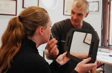
Tear trough filler complications range from minor swelling and lumps to the Tyndall effect and rare vascular events, and most are manageable when identified early and treated by a medically qualified practitioner using hyaluronidase or ultrasound-guided correction.
- The Tyndall effect, caused by filler placed too superficially, produces a bluish tint beneath the skin and is corrected by dissolving the filler with hyaluronidase.
- Persistent swelling beyond two weeks, visible lumps, or asymmetry after tear trough filler warrants a prompt assessment rather than a wait-and-see approach.
- Vascular occlusion is a rare but serious complication requiring immediate medical attention; early recognition of warning signs such as skin colour changes or pain is critical.
- Ultrasound technology supports both the diagnosis of complications and the safe administration of hyaluronidase, helping to confirm filler location before dissolving.
- Most tear trough filler complications are preventable through careful pre-treatment assessment, correct product selection, precise placement technique, and conservative volume.
Tear trough filler complications include the Tyndall effect (a bluish discolouration), persistent swelling, visible lumps, asymmetry, and, in rare cases, vascular occlusion. Most complications are manageable when identified early. Hyaluronic acid-based fillers can be dissolved with hyaluronidase, making prompt assessment by a medically qualified practitioner the most important first step.
Something feels off after your tear trough filler. Maybe there is a bluish tint where you expected a refreshed, rested appearance. Maybe the swelling has not settled the way you were told it would. Or perhaps you have noticed a small lump that was not there before. Whatever has prompted you to read this, you are right to take it seriously and to seek clear information rather than reassurance that everything is probably fine.
Tear trough filler is one of the most technically demanding procedures in non-surgical aesthetic medicine. The anatomy is unforgiving, the margin for error is narrow, and the consequences of poor technique or inappropriate product selection can be visible, uncomfortable, and, in rare cases, medically significant. If you are still in the research phase and have not yet had treatment, the detailed guide to understanding under-eye hollows before considering dermal filler covers the foundational assessment considerations that determine whether filler is appropriate in the first place.
This article focuses specifically on what happens when things do not go as expected: the complications that can arise, how they are identified, how they are managed, and how the risk of encountering them can be meaningfully reduced through careful practitioner selection and assessment-led decision-making.
Tear trough filler complications include the Tyndall effect (a bluish discolouration), persistent swelling, visible lumps, asymmetry, and, in rare cases, vascular occlusion. Most complications are manageable when identified early. Hyaluronic acid-based fillers can be dissolved with hyaluronidase, making prompt assessment by a medically qualified practitioner the most important first step.
—
What complications can occur after tear trough filler, and how common are they?
The under-eye area is one of the least forgiving sites for dermal filler in the face. The skin here is amongst the thinnest on the body, the underlying anatomy is complex, and the tissue has very little capacity to conceal errors in placement, volume, or product choice. Understanding what can go wrong is not meant to alarm you. It is meant to help you recognise when something needs attention.
The Tyndall effect: why filler can turn your under-eye area blue
The Tyndall effect is one of the most commonly discussed complications of tear trough filler, and it is also one of the most visually striking. It occurs when hyaluronic acid filler is placed too superficially within the skin, causing light to scatter as it passes through the gel and reflect back as a bluish or greyish discolouration. The name comes from a physics principle describing how light interacts with particles suspended in a medium, and in aesthetic medicine, it describes exactly what you see when filler sits at the wrong depth beneath very thin skin.
The under-eye area is particularly susceptible because the overlying skin is so fine. Filler that would sit invisibly beneath thicker skin elsewhere on the face can become visible here within days or weeks of treatment. In some cases, the discolouration develops gradually as the filler migrates upwards towards the surface over time.
The Tyndall effect is not a sign of a dangerous complication, but it is a sign of incorrect placement. It does not resolve on its own, and it will not improve with massage. Dissolution with hyaluronidase is the appropriate management, and the discolouration typically clears once the superficially placed filler is removed.
Swelling, persistent oedema, and malar bags: when fluid accumulates around filler
Some degree of swelling after tear trough filler is expected and normal in the first few days. The under-eye area has a rich lymphatic network and tends to swell in response to any injection. What is less expected, and worth understanding, is persistent or delayed oedema that develops or worsens after the initial recovery period.
Hyaluronic acid is hydrophilic, meaning it attracts and retains water. When filler is placed in a tissue plane that is not well-suited to the product, or when the volume used is greater than the tissue can accommodate, the filler can draw additional fluid into the area and create a puffy, swollen appearance that looks worse than the original hollow. This is sometimes referred to as the Oculomotor effect in clinical discussion, where the dynamic movement of the eye and surrounding musculature contributes to fluid accumulation around the filler.
When this fluid settles in the cheek or under-eye area it is known as malar oedema, and it is more likely where the lymphatic drainage in the region is already compromised.
Lumps, nodules, and asymmetry after tear trough filler
Visible lumps or palpable nodules after tear trough filler can have several causes. In some cases, the filler has been placed in a bolus rather than distributed evenly through the tissue. In others, the product has migrated from its original placement. Occasionally, a nodule represents an inflammatory reaction rather than a structural issue with placement.
Asymmetry is also a frequent concern, particularly when one side swells more than the other during recovery or when the volume distribution is uneven. Mild asymmetry in the early weeks may resolve as swelling settles. Persistent asymmetry after six to eight weeks generally requires assessment to determine whether correction or dissolution is appropriate.
—
What is vascular occlusion and why does it matter in the tear trough area?
Vascular occlusion is the most serious complication associated with any injectable filler treatment, and the tear trough area carries a specific risk profile that makes it particularly important to understand. The region is supplied by branches of the ophthalmic artery, which also supplies the eye. In rare cases, filler injected into or near a blood vessel can obstruct blood flow, with consequences that range from localised tissue damage to, in the most severe scenarios, visual compromise.
This is not a complication that occurs frequently when treatment is performed by a medically trained practitioner using appropriate technique and anatomy knowledge. However, it is a complication that demands immediate recognition and action when it does occur, which is why it matters whether your practitioner has the training and emergency management protocols to respond correctly.
Warning signs of vascular compromise you should never ignore
The warning signs of vascular compromise typically appear during or very shortly after injection. Immediate, severe pain that is disproportionate to the procedure is one signal. Skin blanching, a sudden whitening or pallor in the area around the injection site, is another. This blanching may be followed by a dusky, mottled discolouration as the affected tissue becomes ischaemic.
In the tear trough area specifically, any visual disturbance, including blurring, loss of peripheral vision, or sudden changes in how you see, requires emergency management without delay. These symptoms can indicate retrograde embolisation of filler towards the ophthalmic artery, and the window for effective intervention is extremely narrow.
If you experience any of these symptoms during or after treatment at any clinic, do not wait to see whether they resolve. Seek immediate medical assessment.
How vascular occlusion is managed as a medical emergency
Management of vascular occlusion is time-critical. The primary intervention for hyaluronic acid filler occlusion is high-dose hyaluronidase injected promptly into the affected area to dissolve the obstructing filler and restore blood flow. Protocols for managing this complication are well-established in aesthetic medicine literature, and any practitioner performing filler injections should have hyaluronidase immediately available and know how to administer it correctly.
At Dr Alek Nikolic’s practice, ultrasound technology is integrated into the assessment and treatment process. Ultrasound imaging allows visualisation of vascular structures before injection, reducing the risk of inadvertent intravascular placement. It also supports management in the rare event that a complication requires guided intervention. This kind of technology-supported approach reflects the diagnostic-first methodology that underpins how the practice operates.
—
How is tear trough filler dissolved, and what should you expect from the process?
If you are dealing with a complication such as the Tyndall effect, persistent swelling, visible lumps, or asymmetry, dissolution of the existing filler is often the most appropriate first step. Many people feel anxious about this process, partly because it involves another injection into an already sensitive area, and partly because they are uncertain about what it will achieve. Understanding how dissolution works makes the decision easier.
What hyaluronidase does and how it works on hyaluronic acid filler
Hyaluronidase is an enzyme that breaks down hyaluronic acid by cleaving the molecular bonds that give the gel its structure. When injected into or near a deposit of hyaluronic acid filler, it causes the gel to dissolve relatively quickly, typically within 24 to 48 hours for the bulk of the effect, though the tissue continues to remodel over several weeks afterwards.
It is important to understand that hyaluronidase does not discriminate between injected hyaluronic acid and the naturally occurring hyaluronic acid in your own tissue. This means that after dissolution, the area may initially look more hollow than it did before the original filler was placed, particularly if the filler had been present for some time and the surrounding tissue had adapted to it. This is temporary. The body replenishes its natural hyaluronic acid stores over the following weeks, and the appearance typically stabilises before any decision is made about retreatment.
Hyaluronidase is generally well-tolerated. The injection itself causes brief discomfort, similar to any other injection in the area, and topical anaesthetic can be applied beforehand to reduce this.
The role of ultrasound technology in safe filler dissolution
When filler has been placed by another practitioner and its exact location within the tissue is unknown, ultrasound imaging becomes particularly valuable in guiding safe and effective dissolution. Ultrasound allows a practitioner to visualise where the filler sits within the tissue layers, identify any proximity to vascular structures, and direct the hyaluronidase precisely to where it is needed.
This matters for two reasons. First, it reduces the volume of hyaluronidase needed, minimising the effect on surrounding natural tissue. Second, it reduces the risk of inadvertently injecting near a vessel during the dissolution process itself. At Dr Alek Nikolic’s practice, ultrasound-guided assessment is part of how corrective cases are evaluated, particularly when the history of previous treatment is incomplete.
How many sessions does it take to dissolve tear trough filler completely?
The number of dissolution sessions required depends on how much filler is present, how long it has been in place, and which product was used. Older filler that has been present for a year or more may be more integrated into the surrounding tissue and may require more than one treatment. In many cases, a single session achieves the majority of the dissolution, with a follow-up assessment at two to four weeks to evaluate whether further treatment is needed.
It is worth setting realistic expectations: dissolution is not always a single appointment and done. It is a process, and the tissue needs time to settle before the final outcome can be assessed.
—
What makes the tear trough uniquely challenging, and how does assessment reduce complication risk?
Not all filler complications are the result of poor technique in isolation. Many arise from a mismatch between the treatment chosen and the anatomy of the person receiving it. This is why the assessment that precedes any tear trough filler treatment is not a formality. It is the primary determinant of whether the outcome is likely to be safe and satisfying.
Anatomy of the tear trough: why the under-eye area demands precision
The tear trough is a ligamentous structure that separates the lower eyelid from the cheek. Beneath the overlying skin and muscle, the anatomy includes the orbital rim, the pre-periosteal fat, and a network of small but significant blood vessels. The skin overlying this area is amongst the thinnest on the face, with very little subcutaneous fat to buffer the space between the surface and the deeper structures.
This anatomy means that the margin between correct placement and placement that is too superficial, too deep, or too close to a vessel is genuinely narrow. A product that is appropriate for volumising the cheek may be entirely unsuitable for the tear trough because its rheological properties (the way it flows and integrates into tissue) make it too stiff or too prone to water retention in this delicate environment.
Factors such as the prominence of the orbital rim, the degree of skin laxity, the presence of festoons or malar bags, and the depth of the existing hollow all influence whether filler is appropriate and, if so, how it should be placed.
Product selection, placement depth, and volume: the three variables that determine safety
Complications in the tear trough frequently trace back to one or more of three variables: the wrong product, the wrong depth, or too much volume.
Hyaluronic acid-based fillers vary considerably in their cross-linking density, cohesivity, and water-binding capacity. A product with high hydrophilicity placed in the tear trough is likely to attract additional fluid and create or worsen swelling. A product that is too firm will be palpable and potentially visible. The most appropriate products for this area are typically soft, low-cohesivity formulations designed specifically for fine tissue planes.
Placement depth must be periosteal (at the level of the bone) in most cases, keeping the filler away from the superficial skin layers where the Tyndall effect occurs. Volume must be conservative, because the tear trough area has very limited capacity and a small amount of well-placed filler achieves far more than a larger volume placed incorrectly.
—
If you have had tear trough filler elsewhere and are unhappy with the outcome, what are your options?
Arriving at a corrective assessment having had treatment elsewhere is more common than many people realise. The tear trough is a popular treatment site, and the variation in practitioner training, product choice, and injection technique is significant. If you are unhappy with your current outcome, the most important thing to understand is that most complications are addressable, and that an honest assessment is the starting point for any corrective plan.
What a corrective assessment for previous tear trough filler involves
A corrective assessment at Dr Alek Nikolic’s practice begins with a detailed history of your previous treatment, including what product was used if known, when it was placed, and what your concerns are now. This is followed by a careful clinical examination of the under-eye area, assessing the skin quality, the position and volume of any residual filler, the underlying anatomy, and any signs of complications such as the Tyndall effect or asymmetry.
Where the history of previous treatment is unclear or where the filler position is uncertain, ultrasound assessment provides additional information that clinical examination alone cannot offer. This guides the decision about whether dissolution is appropriate, where it should be directed, and what the likely outcome will be.
When dissolving filler is the right first step before any retreatment
In many corrective cases, dissolution before any retreatment is not just appropriate, it is essential. Attempting to add more filler on top of poorly placed existing filler compounds the problem rather than correcting it. The tissue needs to return to a settled baseline before a new treatment plan can be designed with any accuracy.
This can feel frustrating if you are eager to see an improvement, but the clinical rationale is sound. Treating on top of an unknown quantity of existing product carries a higher risk of overfilling, asymmetry, and further complications. Dissolution, followed by a waiting period of four to six weeks, gives both you and your practitioner a clear picture of what you are working with.
Alternative approaches when tear trough filler is not appropriate on reassessment
After dissolution and a period of tissue recovery, reassessment sometimes reveals that tear trough filler is not the most appropriate treatment for your anatomy. This is not a failure of the process. It is the process working correctly.
Some anatomical presentations respond better to cheek volumisation than to direct tear trough treatment, because restoring volume in the mid-face reduces the apparent depth of the hollow without placing any product in the delicate periorbital area. Others may benefit from skin quality improvement through microneedling with Dermapen 4 or skin booster bio-remodelling treatments, which improve the texture and hydration of the overlying skin without adding structural volume. In some cases, the honest assessment is that the degree of skin laxity or the structural anatomy of the lower eyelid makes a surgical opinion the more appropriate referral.
At Dr Alek Nikolic’s practice, the willingness to recommend an alternative or decline retreatment when it is not in your interest is part of what a patient-centred, assessment-led approach means in practice.
—
Frequently asked questions about tear trough filler complications
How do I know if my tear trough filler has been placed too superficially?
The most visible sign is a bluish or greyish discolouration in the under-eye area, which is the Tyndall effect. You may also be able to feel a ridge or lump just beneath the skin surface. If the filler is visible as a slight raised area rather than sitting smoothly within the tissue, this also suggests superficial placement. An assessment by a medically trained practitioner will confirm the diagnosis.
Can tear trough filler cause permanent damage to my eyes or vision?
Permanent visual damage from tear trough filler is extremely rare but not impossible. It is associated with vascular occlusion affecting the ophthalmic artery, which supplies the eye. This risk is significantly reduced when treatment is performed by a practitioner with detailed anatomical knowledge, who uses appropriate technique, and who has hyaluronidase immediately available for emergency management. Prompt recognition and treatment of vascular compromise are critical to preventing permanent harm.
How long does it take for swelling to settle after tear trough filler, and when should I be concerned?
Initial swelling typically settles within one to two weeks. Some residual swelling may persist for up to four weeks, particularly in those who are prone to fluid retention or who had a more reactive response to the injection. Swelling that worsens after the first week, that is significantly asymmetric, or that has not improved at all by four to six weeks warrants an assessment.
Is it safe to dissolve tear trough filler, and does it hurt?
Dissolution with hyaluronidase is generally safe and well-tolerated. The enzyme is specific to hyaluronic acid and works quickly. Topical anaesthetic applied before the injection reduces discomfort significantly. There is a small risk of allergic reaction to hyaluronidase, which a medically trained practitioner will discuss with you beforehand. The process is far less uncomfortable than many people anticipate.
Can I have tear trough filler again after it has been dissolved?
Yes, in most cases. After dissolution, the tissue is allowed to settle for four to six weeks before reassessment. If the anatomy is suitable and the previous complication was related to product choice or placement rather than an inherent unsuitability for filler, retreatment with an appropriate product and technique can achieve a satisfying outcome. The reassessment determines this, not an assumption made in advance.
—
Taking the next step after a tear trough filler complication
If something does not look or feel right after tear trough filler, the worst response is to wait and hope. Most complications are manageable, and the earlier they are assessed, the more straightforward the management tends to be. The Tyndall effect does not resolve without intervention. Persistent swelling does not settle with patience alone. Vascular compromise requires immediate action.
What all of these situations share is that the right next step is an assessment by a medically
Related Articles
- understanding under-eye hollows and whether you are suited to tear trough filler
– The parent pillar covers candidacy assessment, anatomy, and general safety, which provides essential context for readers arriving at this complications article after a treatment has already taken place or while researching risks before proceeding.